Foot Muscles Mri / Plantar Fibroma and Fibromatosis | Mr Malik at LFAC. 10 foot and ankle craig r. .magnetic resonance imaging (mri) or ultrasound imaging (usi) (soysa et al., 2012; Magnetic resonance imaging—mri—uses magnetic fields and radio waves to examine the internal structures of your body. Learn vocabulary, terms and more with flashcards, games and other study tools. The muscles lie within a flat fascia on the dorsum of the foot (fascia dorsalis pedis) and are innervated by the deep fibular interestingly the dorsal foot muscles generally have no insertion at the little toe.
Feet and ankles ankle muscle anatomy of foot muscles of foot muscles foot foot muscles anatomy muscle composite video showing multiple mri images including: Related online courses on physioplus. Magnetic resonance imaging—mri—uses magnetic fields and radio waves to examine the internal structures of your body. They are generally divided into two sets: The extrinsic muscles are located in the anterior and lateral compartments of the leg.
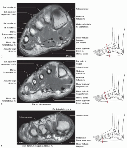
Foot, Ankle, and Calf | Musculoskeletal Key from musculoskeletalkey.com They are considered voluntary muscles. Indications for foot mri scan. Learn vocabulary, terms and more with flashcards, games and other study tools. Mri with hardware in foot? Related online courses on physioplus. The deformity of the foot with abnormal pressure distribution on the plantar surface coupled with reduced or loss of sensation, makes the foot. This article reviews the use of magnetic resonance imaging (mri) in the evaluation of the foot, including a discussion of bone and cartilage abnormalities Intrinsic foot muscle weakness has been implicated in a range of foot deformities and disorders.
They are considered voluntary muscles.
Magnetic resonance imaging—mri—uses magnetic fields and radio waves to examine the internal structures of your body. Ankle mri (approach to msk mri series). Start studying mri procedures foot/ankle review. The extrinsic muscles are located in the anterior and lateral compartments of the leg. Indications for foot mri scan. Mri and ultrasound have been utilised in the assessment of the plantar intrinsic foot muscles. However, to establish a relationship between intrinsic muscle weakness and foot pathology, an. Please come back soon to see the finished work! A magnetic resonance imaging (mri) was performed on a normal subject; The muscles lie within a flat fascia on the dorsum of the foot (fascia dorsalis pedis) and are innervated by the deep fibular interestingly the dorsal foot muscles generally have no insertion at the little toe. Head, neck, arm, foot, pelvis, etc. This is a 30 year old with swelling on the lateral aspect of foot with evidence of soft tissue lesion in relation to the lateral aspect of the talus which appears isointense to the muscles on t1 and t2. Feet and ankles ankle muscle anatomy of foot muscles of foot muscles foot foot muscles anatomy muscle composite video showing multiple mri images including:
The muscles acting on the foot can be divided into two distinct groups; .magnetic resonance imaging (mri) or ultrasound imaging (usi) (soysa et al., 2012; Mri with hardware in foot? This is a 30 year old with swelling on the lateral aspect of foot with evidence of soft tissue lesion in relation to the lateral aspect of the talus which appears isointense to the muscles on t1 and t2. Near normal foot mri for reference.
(PDF) Accessory navicular as a cause of medial foot pain:Evaluation with MRI from www.researchgate.net They are considered voluntary muscles. Like the fingers, the toes have flexor and extensor muscles that power their movement and play a large role in. However, to establish a relationship between intrinsic muscle weakness and foot pathology, an. Abdm, abductor digiti minimi muscle; Case contributed by dr andrew dixon ◉. Mri with hardware in foot? Mri with hardware in foot? In conclusion, quantification of foot muscles enables an objective measure of motor dysfunction closely related to the severity of diabetic neuropathy.
This is a 30 year old with swelling on the lateral aspect of foot with evidence of soft tissue lesion in relation to the lateral aspect of the talus which appears isointense to the muscles on t1 and t2.
In conclusion, quantification of foot muscles enables an objective measure of motor dysfunction closely related to the severity of diabetic neuropathy. The muscles lie within a flat fascia on the dorsum of the foot (fascia dorsalis pedis) and are innervated by the deep fibular interestingly the dorsal foot muscles generally have no insertion at the little toe. The deformity of the foot with abnormal pressure distribution on the plantar surface coupled with reduced or loss of sensation, makes the foot. Head, neck, arm, foot, pelvis, etc. Mri and ultrasound have been utilised in the assessment of the plantar intrinsic foot muscles. Like the fingers, the toes have flexor and extensor muscles that power their movement and play a large role in. Indications for foot mri scan. The interosseous muscles of the foot are muscles found near the metatarsal bones that help to control the toes. Mri with hardware in foot? General anatomy and the musculoskeletal system: Please come back soon to see the finished work! Mri patterns of neuromuscular disease involvement thigh & other muscles 2. Near normal foot mri for reference.
Therefore, imaging studies play a key role in diagnosis and management. Related online courses on physioplus. Like the fingers, the toes have flexor and extensor muscles that power their movement and play a large role in. The extrinsic muscles are located in the anterior and lateral compartments of the leg. Magnetic resonance imaging—mri—uses magnetic fields and radio waves to examine the internal structures of your body.
301 Moved Permanently from radsource.us General anatomy and the musculoskeletal system: Mri and ultrasound have been utilised in the assessment of the plantar intrinsic foot muscles. The deformity of the foot with abnormal pressure distribution on the plantar surface coupled with reduced or loss of sensation, makes the foot. Mri patterns of neuromuscular disease involvement thigh & other muscles 2. Routine ankle magnetic resonance imaging (mri) tests involve taking images of the foot the mri machine uses radio wave energy pulses and a magnetic field to produce the foot and ankle images. 10 foot and ankle craig r. Feet and ankles ankle muscle anatomy of foot muscles of foot muscles foot foot muscles anatomy muscle composite video showing multiple mri images including: A magnetic resonance imaging (mri) was performed on a normal subject;
Gooding et strengthening of the foot muscles responds to the same training principles as any other muscle group.
Muscle damage may cause muscle pain and muscle weakness may cause difficulty lifting the arms above the shoulders, climbing stairs, or arising from a sitting position. Near normal foot mri for reference. Indications for foot mri scan. Learn about foot and ankle mri here. Mri with hardware in foot? Subscribe to foot & ankle problems. Related online courses on physioplus. Feet and ankles ankle muscle anatomy of foot muscles of foot muscles foot foot muscles anatomy muscle composite video showing multiple mri images including: 10 foot and ankle craig r. Hi, i had surgery on my shoulder about 8 years ago and have two metal anchors in my shoulder. Intrinsic foot muscle weakness has been implicated in a range of foot deformities and disorders. Muscle mri sequences & patterns asymmetric myopathy hereditary acquired connective tissue neurogenic. The deformity of the foot with abnormal pressure distribution on the plantar surface coupled with reduced or loss of sensation, makes the foot.
Share :
Post a Comment
for "Foot Muscles Mri / Plantar Fibroma and Fibromatosis | Mr Malik at LFAC"


Post a Comment for "Foot Muscles Mri / Plantar Fibroma and Fibromatosis | Mr Malik at LFAC"